CT Scans
Surgical Microscopes

There are cases that would have almost certainly required extraction just a few years ago and now can be salvaged by the benefits of the magnification and illumination the microscope provides. Benefits of using a surgical microscope are removal of separated instruments, locating missed or blocked canals and visualizing and determining the extent of cracks or fractures.
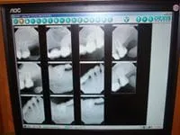
In addition, images can be captured through the microscope and viewed for better understanding. We also utilize digital radiography not only for the high reduction in x-ray exposure, but for the benefits it provides for explanations. A large radiographic image, when seen on the computer monitor, becomes an invaluable aid in communication.
Digital Radiography


